Cyberonics reneged on its "Lifetime Reimbursement Guarantee". Click on the image to learn how you can help...

Tuesday, September 30, 2014

Transcranial Magnetic Stimulation Offers Hope for Treatment-Resistant Depression

Therese Borchard
Sanity Break

Help for depression and anxiety


Transcranial Magnetic Stimulation Offers Hope for Treatment-Resistant Depression

By Therese Borchard

Published Sep 29, 2014

In December of 2012, Stephanie S. was taking 300 mgs of sertraline (Zoloft), 300 mgs of (bupropion Hcl) Wellbutrin, 300 mgs of trazodone hydrochloride (Desyrel), 200 mgs of risperidone (Risperdal), and 8 mgs of alprazolam (Xanax), but was as depressed as she has ever been. She had also gained 100 pounds as a side effect of all the medications. Having tried a total of 10 different kinds of drugs with no success, her doctor recommended transcranial magnetic stimulation (TMS), a non-invasive procedure that stimulates nerve cells in the brain with short magnetic pulses. A large electromagnetic coil is placed against the scalp which generates focused pulses that pass through the skull and stimulate the cerebral cortex of the brain, a region that regulates mood. The procedure was approved by the FDA in 2008.

She didn’t feel any difference after 11 treatments, but she can vividly remember the morning after her 12th treatment. She explained:

I woke up… I mean WOKE UP!! I felt so light, instead of feeling weighed down. The sun was brighter. My overall feeling was JOY. This was unfamiliar to me, and I loved it. I came downstairs grinning from ear to ear and just looked at my husband. He knew! I just threw my arms around his neck and laughed. The feeling was indescribable. It was NIRVANA!! I felt better than I had felt before my breakdown. It was MAGICAL! I think that was the first time in my life that I felt pure joy!

She continued and finished the 30 treatments.

Dr. Kira Stein, MD, board certified psychiatrist and medical director of West Coast TMS Institute in Sherman Oaks, Los Angeles, is excited about the success she’s had in treating her patients with TMS. She usually does five sessions a week, for a total of 30 sessions; the entire procedure lasts about six to eight weeks, though some patients may need more treatment to respond.

She estimates that about one-third of TMS patients have a full remission and no longer experience depression symptoms.

One-half of TMS patients respond signficantly, where their depression symptoms are improved by at least 50 percent, but do not reach complete remission. The more depressive cycles they have had in their lives, the more difficult it is to treat them in general.

Dr. Stein’s experience is that TMS success rates are higher when TMS is used as an augmentation strategy for patients who have only partially responded to medication, or who cannot tolerate higher medication doses. She usually recommends a person stay on a dose of maintenance antidepressants and finds that some patients need maintenance TMS treatments to stay well.

The treatment is expensive. Dr. Stein says that each session (and on average people usually require 30) run from $300 to $450, a session; however, more and more insurance companies are picking up at least some of the bill.

Stephanie paid $7,450 out of pocket. Her insurance chipped in $7,000 (the total cost was about $14,000).

Stephanie stayed in remission for a year and a half until a cascade of tragic events, including the suicide of her sister, caused a relapse of depressive symptoms. When different kinds of medication again did little to relieve her pain, she decided to do TMS for a second time.

She’s been participating in the online depression support group I moderate on Facebook. About a month ago, I remember a distinct change in the tone of her posts. They went from being desperate to hopeful, from cynical to curious, and from flat to playful.

“What’s the matter with me?” she asked the group. “On the way to my husband’s work, I’m noticing everything for the first time.”

“I think your TMS treatment is working,” I replied.

“Yes!” she said. “I laughed again!!”

She has eight treatments left, and hopes she continues to laugh for a very long time.

Posted in: Depression

6 Comments

http://www.everydayhealth.com/columns/therese-borchard-sanity-break/transcranial-magnetic-stimulation-tms-offers-hope-for-treatment-resistant-depression/

New study reports how TMS treatment works in people with depression

New study reports how TMS treatment works in people with depression

Published on September 30, 2014 at 12:21 PM · No Comments


A new study in Biological Psychiatry reports how magnetic stimulation treatment works

On Star Trek, it is easy to take for granted the incredible ability of futuristic doctors to wave small devices over the heads of both humans and aliens, diagnose their problems through evaluating changes in brain activity or chemistry, and then treat behavior problems by selectively stimulating relevant brain circuits.

While that day is a long way off, transcranial magnetic stimulation (TMS) of the left dorsolateral prefrontal cortex does treat symptoms of depression in humans by placing a relatively small device on a person's scalp and stimulating brain circuits. However, relatively little is known about how, exactly, TMS produces these beneficial effects.

Some studies have suggested that TMS may modulate atypical interactions between two large-scale neuronal networks, the frontoparietal central executive network (CEN) and the medial prefrontal-medial parietal default mode network (DMN). These two functional networks play important roles in emotion regulation and cognition.

In order to advance our understanding of the underlying antidepressant mechanisms of TMS, Drs. Conor Liston, Marc Dubin, and their colleagues conducted a longitudinal study to test this hypothesis.

The researchers used functional magnetic resonance imaging in 17 currently depressed patients to measure connectivity in the CEN and DMN networks both before and after a 25-day course of TMS. They also compared the connectivity in the depressed patients with a group of 35 healthy volunteers.

Related Stories

TMS normalized depression-related hyperconnectivity between the subgenual cingulate and medial prefrontal areas of the DMN, but did not alter connectivity in the CEN.

Liston, an Assistant Professor at Weill Cornell Medical College, further details their findings, "We found that connectivity within the DMN and between nodes of the DMN and CEN was elevated in depressed individuals compared to healthy volunteers at baseline and normalized after TMS. Additionally, individuals with greater baseline connectivity with subgenual anterior cingulate cortex - an important target for other antidepressant modalities - were more likely to respond to TMS."

These findings indicate that TMS may act, in part, by selectively regulating network-level connectivity.

Dr. John Krystal, Editor of Biological Psychiatry, comments, "We are a long way from Star Trek, but even the current ability to link brain stimulation treatments for depression to the activity of particular brain circuits strikes me as incredible progress."

Dubin, also an Assistant Professor at Weill Cornell Medical College, adds, "Our findings may inform future efforts to develop personalized strategies for treating depression with TMS based on the connectivity of an individual's default mode network. Further, they may help triage to TMS only those patients most likely to respond."

http://www.news-medical.net/news/20140930/New-study-reports-how-TMS-treatment-works-in-people-with-depression.aspx

Tuesday, September 23, 2014

Safety of a dedicated brain MRI protocol in patients with a vagus nerve stimulator.

Epilepsia. 2014 Sep 19. doi: 10.1111/epi.12774. [Epub ahead of print]

Safety of a dedicated brain MRI protocol in patients with a vagus nerve stimulator.

de Jonge JC1, Melis GI, Gebbink TA, de Kort GA, Leijten FS.

Author information
  • 1Department of Neurology and Neurosurgery, Brain Center Rudolf Magnus, University Medical Center Utrecht, Utrecht, The Netherlands.
Abstract

Although implanted metallic devices constitute a relative contraindication to magnetic resonance imaging (MRI) scanning, the safety of brain imaging in a patient with a vagus nerve stimulator (VNS) is classified as "conditional," provided that specific manufacturer guidelines are followed when a transmit and receive head coil is used at 1.5 or 3.0 Tesla. The aim of this study was to evaluate the safety of performing brain MRI scans in patients with the VNS. From September 2009 until November 2011, 101 scans were requested in 73 patients with the VNS in The Netherlands. Patients were scanned according to the manufacturer's guidelines. No patient reported any side effect, discomfort, or pain during or after the MRI scan. In one patient, a lead break was detected based on device diagnostics after the MRI-scan. However, because no system diagnostics had been performed prior to MR scanning in this patient, it is unclear whether MR scanning was responsible for the lead break. The indication for most scans was epilepsy related. Twenty-six scans (26%) were part of a (new) presurgical evaluation and could probably better have been performed prior to VNS implantation. Performing brain MRI scans in patients with an implanted VNS is safe when a modified MRI protocol is followed.

Wiley Periodicals, Inc. © 2014 International League Against Epilepsy.

KEYWORDS:

Epilepsy; Magnetic resonance imaging; Patient safety; Vagus nerve stimulator

PMID:
25244102
[PubMed - as supplied by publisher] http://www.ncbi.nlm.nih.gov/pubmed/25244102
Related citations in PubMed

See reviews...See all...

Monday, September 22, 2014

Evidence supports deep brain stimulation for obsessive-compulsive disorder

PUBLIC RELEASE DATE:

22-Sep-2014

[ Print | E-mail ] Share Share

[ Close Window ]
Contact: Connie Hughes
Connie.Hughes@wolterskluwer.com
646-674-6348
Wolters Kluwer Health
@WKHealth

Evidence supports deep brain stimulation for obsessive-compulsive disorder

Bilateral stimulation is effective for OCD that doesn't respond to medications, says new guideline in Neurosurgery

September 22, 2014 – Available research evidence supports the use of deep brain stimulation (DBS) for patients with obsessive-compulsive disorder (OCD) who don't respond to other treatments, concludes a review in the October issue of Neurosurgery, official journal of the Congress of Neurological Surgeons (CNS). The journal is published by Lippincott Williams & Wilkins, a part of Wolters Kluwer Health.

Based on evidence, two specific bilateral DBS techniques are recommended for treatment of carefully selected patients with OCD, according to a new clinical practice guideline endorsed by the CNS and the American Association of Neurological Surgeons. While calling for further research in key areas, Dr. Clement Hamani of Toronto Western Hospital and coauthors emphasize that patients with OCD symptoms that don't respond to other treatments should continue to have access to DBS.

Deep Brain Stimulation for OCD—What's the Evidence?

Dr. Hamani led a multispecialty expert group in performing a systematic review of research on the effectiveness of DBS for OCD. Deep brain stimulation—placement of electrodes in specific areas of the brain, followed by electrical stimulation of those areas—has become an important treatment for patients with Parkinson's disease and other movement disorders.

Although many patients with OCD respond well to medications and/or psychotherapy, 40 to 60 percent continue to experience symptoms despite treatment. Over the past decade, a growing number of reports have suggested that DBS may be an effective alternative in these "medically refractory" cases.

Dr. Hamani and colleagues were tasked with analyzing the supporting evidence and developing an initial clinical practice guideline for the use of DBS for patients with OCD. The review and guideline development process was sponsored by the American Society of Stereotactic and Functional Neurosurgery and the CNS. Out of more than 350 papers, the reviewers identified seven high-quality studies evaluating DBS for OCD.

Based on that evidence, they conclude that bilateral stimulation (on both sides of the brain) of two brain "targets"—areas called the subthalamic nucleus and the nucleus accumbens—can be regarded as effective treatments for OCD. In controlled clinical trials, both techniques improved OCD symptoms by around 30 percent on a standard rating scale.

While Research Proceeds, well-selected treatment-resistant severe OCD Patients Should Have Access to DBS

That evidence forms the basis for a clinical guideline stating that bilateral DBS is a "reasonable therapeutic option" for patients with severe OCD that does not respond to other treatments. The guideline also notes that there is "insufficient evidence" supporting the use of any type of unilateral DBS target (one side of the brain) for OCD.

The review highlights the difficulties of studying the effectiveness of DBS for OCD—because most patients respond to medical treatment, studies of this highly specialized treatment typically include only small numbers of patients. Dr. Hamani and coauthors identify some priorities for future research: particularly to identify the most effective brain targets and the subgroups of patients most likely to benefit.

Despite the limited evidence base, DBS therapy for OCD has been approved by the Food and Drug Administration under a humanitarian device exemption. Dr. Hamani and coauthors note that various safeguards are in place to ensure appropriate use, and prevent overuse, of DBS for OCD.

While research continues, they believe that functional neurosurgeons should continue to work with other specialists to ensure that patients with severe, medically refractory OCD continue to have access to potentially beneficial DBS therapy.

###

Click here to read "Deep Brain Stimulation for Obsessive-Compulsive Disorder: Systematic Review and Evidence-Based Guideline Sponsored by the American Society for Stereotactic and Functional Neurosurgery and the Congress of Neurological Surgeons (CNS) and Endorsed by the CNS and American Association of Neurological Surgeons."

About Neurosurgery

Neurosurgery, the Official Journal of the Congress of Neurological Surgeons, is your most complete window to the contemporary field of neurosurgery. Members of the Congress and non-member subscribers receive 3,000 pages per year packed with the very latest science, technology, and medicine, not to mention full-text online access to the world's most complete, up-to-the-minute neurosurgery resource. For professionals aware of the rapid pace of developments in the field, Neurosurgery is nothing short of indispensable.

About Wolters Kluwer Health

Wolters Kluwer Health is a leading global provider of information, business intelligence and point-of-care solutions for the healthcare industry. Serving more than 150 countries worldwide, clinicians rely on Wolters Kluwer Health's market leading information-enabled tools and software solutions throughout their professional careers from training to research to practice. Major brands include Health Language®, Lexicomp®, Lippincott Williams & Wilkins, Medicom®, Medknow, Ovid®, Pharmacy OneSource®, ProVation® Medical and UpToDate®.

Wolters Kluwer Health is part of Wolters Kluwer, a market-leading global information services company. Wolters Kluwer had 2013 annual revenues of €3.6 billion ($4.7 billion), employs approximately 19,000 people worldwide, and maintains operations in over 40 countries across Europe, North America, Asia Pacific, and Latin America.maintains operations in over 40 countries across Europe, North America, Asia Pacific, and Latin America. Wolters Kluwer is headquartered in Alphen aan den Rijn, the Netherlands. Its shares are quoted on Euronext Amsterdam (WKL) and are included in the AEX and Euronext 100 indices. Wolters Kluwer has a sponsored Level 1 American Depositary Receipt program. The ADRs are traded on the over-the-counter market in the U.S. (WTKWY).

Follow our official Twitter handle: @WKHealth.

http://www.eurekalert.org/pub_releases/2014-09/wkh-esd092214.php

Friday, September 19, 2014

Vagus nerve stimulation for drug-resistant epilepsy: A European long-term study up to 24 months in 347 children.

Epilepsia. 2014 Sep 17. doi: 10.1111/epi.12762. [Epub ahead of print]

Vagus nerve stimulation for drug-resistant epilepsy: A European long-term study up to 24 months in 347 children.

Orosz I1, McCormick D, Zamponi N, Varadkar S, Feucht M, Parain D, Griens R, Vallée L, Boon P, Rittey C, Jayewardene AK, Bunker M, Arzimanoglou A, Lagae L.

Author information
  • 1Department of Neuropediatrics, Children's Hospital, University of Leubeck, Leubeck, Germany.
Abstract
OBJECTIVE:

To gain insight into the long-term impact of vagus nerve stimulation (with VNS Therapy) in children with drug-resistant epilepsy, we conducted the largest retrospective multicenter study to date over an extended follow-up period of up to 24 months.

METHODS:

The primary objective was to assess change in seizure frequency of the predominant seizure type (defined as the most disabling seizure) following VNS device implantation. Treating physicians collected data from patient records from baseline to 6, 12, and 24 months of follow-up.

RESULTS:

The analysis population included 347 children (aged 6 months to 17.9 years at the time of implant). At 6, 12, and 24 months after implantation, 32.5%, 37.6%, and 43.8%, respectively, of patients had ≥50% reduction in baseline seizure frequency of the predominant seizure type. The responder rate was higher in a subgroup of patients who had no change in antiepileptic drugs (AEDs) during the study. Favorable results were also evident for all secondary outcome measures including changes in seizure duration, ictal severity, postictal severity, quality of life, clinical global impression of improvement, and safety. Post hoc analyses demonstrated a statistically significant correlation between VNS total charge delivered per day and an increase in response rate. VNS Therapy is indicated as adjunctive therapy in children with focal, structural epilepsies, who for any reason are not good candidates for surgical treatment following the trial of two or more AEDs. Children with predominantly generalized seizures from genetic, structural epilepsies, like Dravet syndrome or Lennox-Gastaut syndrome, could also benefit from VNS Therapy.

SIGNIFICANCE:

The results demonstrate that adjunctive VNS Therapy in children with drug-resistant epilepsy reduces seizure frequency and is well tolerated over a 2-year follow-up period. No new safety issues were identified. A post hoc analysis revealed a dose-response correlation for VNS in patients with epilepsy.

Wiley Periodicals, Inc. © 2014 International League Against Epilepsy.

KEYWORDS:

Clinical trial; Epilepsy; Pediatrics; Quality of life; Vagus nerve stimulation

PMID:
25231724
[PubMed - as supplied by publisher] http://www.ncbi.nlm.nih.gov/pubmed/25231724

Wednesday, September 17, 2014

Response to TMS Sustained in Depression

Medscape Medical News > Psychiatry

Response to TMS Sustained in Depression

Pam Harrison

September 17, 2014


The final analysis of a study evaluating transcranial magnetic stimulation (TMS) confirms that the majority of patients with resistant depression who respond to TMS in the acute setting continue to have a sustained response for a period of 1 year, an observational trial shows.

David Dunner, MD, director, Center for Anxiety and Depression, Mercer Island, Washington, and colleagues reported that 68% of 257 patients who had received TMS during the acute treatment phase had improved by the end of the 12-month follow-up and that 45% had experienced complete remission at 1 year.

"These are patients with treatment-resistant depression who are difficult to treat, who generally do not get better easily. TMS not only got people better with the usual course of about 30 treatments in the acute treatment phase, but we showed that the benefit was maintained over a 1-year period in the majority of patients who improved," Dr. Dunner told Medscape Medical News.

Courtesy of Neuronetics, Inc.

"So I view TMS as a treatment for people who have a new depression and who have a history of failing 3 or 4 prior treatments for depression, where it is unlikely that the antidepressant is going to work. In that population, I think we get good results, and the results justify the use of TMS and the cost of treatment as well."

The final results of the observational study were published online on September 16 in the Journal of Clinical Psychiatry. Preliminary long-term results were first reported from the American Psychiatric Association meeting in 2013 by Medscape Medical News. The study was supported by Neuronetics Inc.

Long-term Results

As investigators previously reported ( Depress Anxiety. 2012;29:587-596), 62% of patients had achieved symptomatic improvement, and 41% had achieved a complete remission by the end of the acute treatment phase.

http://www.medscape.com/viewarticle/831860

Wednesday, September 10, 2014

It Ain't Necessarily So: Why Much of the Medical Literature Is Wrong

It Ain't Necessarily So: Why Much of the Medical Literature Is Wrong

Christopher Labos, MD CM, MSc, FRCPC

September 09, 2014

17 comments

In 1897, eight-year-old Virginia O'Hanlon wrote to the New York Sun to ask, "Is there a Santa Claus?"[1] Virginia's father, Dr. Phillip O'Hanlon, suggested that course of action because "if you see it in the Sun, it's so." Today many clinicians and health professionals may share the same faith in the printed word and assume that if it says it in the New England Journal of Medicine (NEJM) or JAMA or The Lancet, then it's so.

Putting the existence of Santa Claus aside, John Ioannidis[2] and others have argued that much of the medical literature is prone to bias and is, in fact, wrong.

Given a statistical association between X and Y, most people make the assumption that X caused Y. However, we can easily come up with 5 other scenarios to explain the same situation.

1. Reverse Causality

Given the association between X and Y, it is actually equally likely that Y caused X as it is that X caused Y. In most cases, it is obvious which variable is the cause and which is the effect. If a study showed a statistical association between smoking and coronary heart disease (CHD), it would be clear that smoking causes CHD and not that CHD makes people smoke. Because smoking preceded CHD, reverse causality in this case is impossible. But the situation is not always that clear-cut. Consider a study published in the NEJM that showed an association between diabetes and pancreatic cancer.[3] The casual reader might conclude that diabetes causes pancreatic cancer. However, further analysis showed that much of the diabetes was of recent onset. The pancreatic cancer preceded the diabetes, and the cancer subsequently destroyed the insulin-producing islet cells of the pancreas. Therefore, this was not a case of diabetes causing pancreatic cancer but of pancreatic cancer causing the diabetes.

Mistaking what came first in the order of causation is a form of protopathic bias.[4] There are numerous examples in the literature. For example, an assumed association between breast feeding and stunted growth, [5] actually reflected the fact that sicker infants were preferentially breastfed for longer periods. Thus, stunted growth led to more breastfeeding, not the other way around. Similarly, an apparent association between oral estrogens and endometrial cancer was not quite what it seemed.[6] Oral estrogens may be prescribed for uterine bleeding, and the bleeding may be caused by an undiagnosed cancer. Therefore, when the cancer is ultimately diagnosed down the road, it will seem as if the estrogens came before the cancer, when in fact it was the cancer (and the bleeding) that led to the prescription of estrogens. Clearly, sometimes it is difficult to disentangle which factor is the cause and which is the effect.

2. The Play of Chance and the DICE Miracle

Whenever a study finds an association between 2 variables, X and Y, there is always the possibility that the association was simply the result of random chance.

Most people assess whether a finding is due to chance by checking if the P value is less than .05. There are many reasons why this the wrong way to approach the problem, and an excellent review by Steven Goodman[7] about the popular misconceptions surrounding the P value is a must-read for any consumer of medical literature.

To illustrate the point, consider the ISIS-2 trial,[8] which showed reduced mortality in patients given aspirin after myocardial infarction. However, subgroup analyses identified some patients who did not benefit: those born under the astrological signs of Gemini and Libra; patients born under other zodiac signs derived a clear benefit with a P value < .00001. Unless we are prepared to re-examine the validity of astrology, we would have to admit that this was a spurious finding due solely to chance. Similarly, Counsell et al. performed an elegant experiment using 3 different colored dice to simulate the outcomes of theoretical clinical trials and subsequent meta-analysis.[9] performed an elegant experiment using 3 different colored dice to simulate the outcomes of theoretical clinical trials and subsequent meta-analysis. Students were asked to roll pairs of dice, with a 6 counting as patient death and any other number correlating to survival. The students were told that one dice may be more "effective" or less effective (ie, generate more sixes or study deaths). Sure enough, no effect was seen for red dice, but a subgroup of white and green dice showed a 39% risk reduction (P = .02). Some students even reported that their dice were "loaded." This finding was very surprising because Counsell had played a trick on his students and used only ordinary dice. Any difference seen for white and green dice was a completely random result.

The Frequency of False Positives

It is sometimes humbling and fairly disquieting to think that chance can play such a large role in the results of our analyses. Subgroup analyses, as shown above, are particularly prone to spurious associations. Most researchers set their significance level or rate of type 1 error at 5%. However, if you perform 2 analyses, then the chance of at least one of these tests being "wrong" is 9.75%. Perform 5 tests, and the probability becomes 22.62%; and with 10 tests, there is a 40.13% of at least 1 spurious association even if none of them are actually true. Because most papers present many different subgroups and composite endpoints, the chance of at least one spurious association is very high. Often, the one spurious association is published, and the other negative tests never see the light of day.[10]

There is a way to guard against such spurious findings: replication. Unfortunately, the current structure of academic medicine does not favor the replication of published results,[11] and several studies have shown that many published trials do not stand up to independent verification and are likely false positives.[12,13] In 2005, John Ioannidis published a review of 45 highlighted studies in major medical journals. He found that 24% were never replicated, 16% were contradicted by subsequent research, and another 16% were shown to have smaller effect sizes than originally reported. Less than half (44%) were truly replicated.

The frequency of these false-positive studies in the published literature can be estimated to some degree.[2] Consider a situation in which 10% of all hypotheses are actually true. Now consider that most studies have a type 1 error rate (the probability of claiming an association when none exists [ie, a false positive]) of 5% and a type 2 error rate (the probability of claiming there is no association when one actually exists [ie, a false negative)] of 20%, which are the standard error rates presumed by most clinical trials. This allows us to create the following 2x2 table.

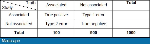
By plugging in the numbers above:

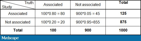
This would imply that of the 125 studies with a positive finding, only 80/125 or 64% are true. Therefore, one third of statistically significant findings are false positives purely by random chance. That assumes, of course, that there is no bias in the studies, which we will deal with presently.

3. Bias: Coffee, Cellphones, and Chocolate

Bias occurs when there is no real association between X and Y, but one is manufactured because of the way we conducted our study. Delgado-Rodriguez and Llorca[4] identified 74 types of bias in their glossary of the most common biases, which can be broadly categorized into 2 main types: selection bias and information bias.

One classic example of selection bias occurred in 1981 with a NEJM study showing an association between coffee consumption and pancreatic cancer.[15] The selection bias occurred when the controls were recruited for the study. The control group had a high incidence of peptic ulcer disease, and so as not to worsen their symptoms, they drank little coffee. Thus, the association between coffee and cancer was artificially created because the control group was fundamentally different from the general population in terms of their coffee consumption. When the study was repeated with proper controls, no effect was seen.[16]

Information bias, as opposed to selection bias, occurs when there is a systematic error in how the data are collected or measured. Misclassification bias occurs when the measurement of an exposure or outcome is imperfect; for example, smokers who identify themselves as nonsmokers to investigators or individuals who systematically underreport their weight or overreport their height.[17] A special situation, known as recall bias, occurs when subjects with a disease are more likely to remember the exposure under investigation than controls. In the INTERPHONE study, which was designed to investigate the association between cell phones and brain tumors, a spot-check of mobile phone records for cases and controls showed that random recall errors were large for both groups with an overestimation among cases for more distant time periods.[18] Such differential recall could induce an association between cell phones and brain tumors even if none actually exists.

An interesting type of information bias is the ecological fallacy. The ecological fallacy is the mistaken belief that population-level exposures can be used to draw conclusions about individual patient risks.[4] A recent example of the ecological fallacy, was a tongue-in-cheek NEJM study by Messerli[19} showing that countries with high chocolate consumption won more Nobel prizes. The problem with country-level data is that countries don't eat chocolate, and countries don't win Nobel prizes. People eat chocolate, and people win Nobel prizes. This study, while amusing to read, did not establish the fundamental point that the individuals who won the Nobel prizes were the ones actually eating the chocolate.[20]

Another common ecological fallacy is the association between height and mortality. There are a number of reviews suggesting that shorter stature is associated with a longer life span.[21] However, most of these studies looked at country-level data. Danes are taller than Italians and also have more coronary heart disease. However, if you look at twins[22] or individuals within the same country,[23] you see the opposite association -- namely, it is the shorter individuals who have more heart disease. Again, the fault lies in looking at countries rather than individuals.

4. Confounding

Confounding, unlike bias, occurs when there really is an association between X and Y, but the magnitude of that association is influenced by a third variable. Whereas bias is a human creation, the product of inappropriate patient selection or errors in data collection, confounding exists in nature.[24]

For example, diabetes confounds the relationship between renal failure and heart disease because it can lead to both conditions. Although patients with renal failure are at higher risk for heart disease, failing to account for the inherent risk of diabetes makes that association seem stronger than it actually is.

Confounding is a problem in every observational study, and statistical adjustment cannot always eliminate it. Even some of the best observational trials fall victim to confounding. Hormone replacement therapy was long thought to be protective for cardiac disease[25] until the Women’s Health Initiative randomized trial refuted that notion.[26] Despite the best attempts at statistical adjustment, there can always be residual confounding. However, simply putting more variables into a multivariate model is not necessarily a better option. Overadjusting can be just as problematic, and adjusting for unnecessary variables can lead to biased results.[27,28]

Real-World Randomization

Confounding can be dealt with through randomization. When study subjects are randomly allocated to one group or another purely by chance, any confounders (even unknown confounders) should be equally present in both the study and control group. However, that assumes that randomization was handled correctly. A 1996 study sought to compare laparoscopic vs open appendectomy for appendicitis.[29] The study worked well during the day, but at night the presence of the attending surgeon was required for the laparoscopic cases but not the open cases. Consequently, the on-call residents, who didn't like calling in their attendings, adopted a practice of holding the translucent study envelopes up to the light to see if the person was randomly assigned to open or laparoscopic surgery. When they found an envelope that allocated a patient to the open procedure (which would not require calling in the attending and would therefore save time), they opened that envelope and left the remaining laparoscopic envelopes for the following morning. Because cases operated on at night were presumably sicker than those that could wait until morning, the actions of the on-call team biased the results. Sicker cases preferentially got open surgery, making the outcomes of the open procedure look worse than they actually were.[30] So, though randomized trials are often thought of as the solution to confounding, if randomization is not handled properly, confounding can still occur. In this case, an opaque envelope would have solved the problem.

5. Exaggerated Risk

Finally, let us make the unlikely assumption that we have a trial where nothing went wrong, and we are free of all of the problems discussed above. The greatest danger lies in our misinterpretation of the findings. A report in the New England Journal of Medicine reported that African Americans were 40% less likely to be sent for an angiogram than their white counterparts.[31] The report generated considerable media attention at the time, but a later article by Schwartz et al.[32] pointed out that the results were overstated. Had the authors used a risk ratio instead of an odds ratio, the result would have been 7% instead of 40%, and it's unlikely that the paper would have been given such prominence. Choosing the correct statistical test can be difficult. Nearly 20 years ago. Sackett and colleagues[33] proclaimed "Down with odds ratios!"[33] and yet they remain frequently used in the literature.

Another major problem is the use of relative risks vs absolute risks. Although the latter are clearly preferable, one review of almost 350 studies found that 88% never reported the absolute risk.[34] Furthermore, overreliance on relative risks can be very misleading. Baylin and colleagues[35] reported that the relative risk for myocardial infarction in the hour after drinking a cup of coffee was 1.5 (ie, a 50% increase). This rather concerning finding was taken up by Poole in a bitingly satirical letter to the editor,[36] in a bitingly satirical letter to the editor, where he calculated that the relative risk of 1.5 translated to an absolute risk of 1 heart attack for every 2 million cups of coffee. Clearly, well-done studies have to be put in clinical context, and it is paramount to remember that statistical significance does not imply clinical significance.

Why Bother?

With all of the different ways that clinical trials can go wrong, one might wonder why we bother at all. Unlike little Virginia, who was prepared to believe whatever she saw in the newspaper, we have become, if not cynics, then at least skeptics when it comes to our published research. But skepticism is a good thing and makes us challenge what we think we know in favor of what we can prove. Without this skepticism, we would still be prescribing hormone replacement therapy to prevent heart disease in women, giving class I anti-arrhythmics to cardiac patients after myocardial infarction, and prescribing COX-2 inhibitors with reckless abandon.

As Dr. Fiona Godlee summed up in her BMJ editorial on evidence-based medicine, “[it’s a] flawed system but still the best we’ve got.”[37]

References
  1. Berman R. Virginia O'Hanlon's home to be turned into school. December 16, 2005. http://www.nysun.com/new-york/virginia-ohanlons-home-to-be-turned-into-school/24556 Accessed July 24, 2014.

  2. Ioannidis JP. Why most published research findings are false. PLoS Med. 2005;2:e124.

  3. Gullo L, Pezzilli R, Morselli-Labate AM; Italian Pancreatic Cancer Study Group. Diabetes and the risk of pancreatic cancer. N Engl J Med. 1994;331:81-84. Abstract

  4. Delgado-RodrĂ­guez M, Llorca J. Bias. J Epidemiol Community Health. 2004;58:635-641. Abstract

  5. Marquis GS, Habicht JP, Lanata CF, Black RE, Rasmussen KM. Association of breastfeeding and stunting in Peruvian toddlers: an example of reverse causality. Int J Epidemiol. 1997;26:349-356. Abstract

  6. Horwitz RI, Feinstein AR. Analysis of clinical susceptibility bias in case-control studies. Analysis as illustrated by the menopausal syndrome and the risk of endometrial cancer. Arch Intern Med. 1979;139:1111-1113. Abstract

  7. Goodman S. A dirty dozen: twelve P-value misconceptions. Semin Hematol. 2008;45:135-140. Abstract

  8. ISIS-2 (Second International Study of Infarct Survival) Collaborative Group. Randomised trial of intravenous streptokinase, oral aspirin, both, or neither among 17,187 cases of suspected acute myocardial infarction: ISIS-2. Lancet. 1988;332:349-360.

  9. Counsell CE, Clarke MJ, Slattery J, Sandercock PA. The miracle of DICE therapy for acute stroke: fact or fictional product of subgroup analysis? BMJ. 1994;309:1677-1681. Abstract

  10. Dwan K, Altman DG, Arnaiz JA, et al. Systematic review of the empirical evidence of study publication bias and outcome reporting bias. PLoS One. 2008;3:e3081.

  11. Hartshorne JK, Schachner A. Tracking replicability as a method of post-publication open evaluation. Front Comput Neurosci. 2012;6:8.

  12. Boffetta P, McLaughlin JK, La Vecchia C, Tarone RE, Lipworth L, Blot WJ. False-positive results in cancer epidemiology: a plea for epistemological modesty. J Natl Cancer Inst. 2008;100:988-995. Abstract

  13. Ioannidis JP, Ntzani EE, Trikalinos TA, Contopoulos-Ioannidis DG. Replication validity of genetic association studies. Nat Genet. 2001;29:306-309. Abstract

  14. Ioannidis JP. Contradicted and initially stronger effects in highly cited clinical research. JAMA. 2005;294:218-228. Abstract

  15. MacMahon B, Yen S, Trichopoulos D, Warren K, Nardi G. Coffee and cancer of the pancreas. N Engl J Med. 1981;304:630-633. Abstract

  16. Gold EB, Gordis L, Diener MD, et al. Diet and other risk factors for cancer of the pancreas. Cancer. 1985;55:460-467.

  17. Shields M, Connor Gorber S, Janssen I, Tremblay MS. Bias in self-reported estimates of obesity in Canadian health surveys: an update on correction equations for adults. Health Rep. 2011;22:35-45. Abstract

  18. Vrijheid M, Armstrong BK, Bedard D, et al. Recall bias in the assessment of exposure to mobile phones. J Expo Sci Environ Epidemiol. 2009;19:369-381. Abstract

  19. Messerli FH. Chocolate consumption, cognitive function, and Nobel laureates. N Engl J Med. 2012;367:1562-1564. Abstract

  20. Dunstan F. Nobel prizes, chocolate and milk: the statistical view. Pract Neurol. 2013;13:206-207. Abstract

  21. Samaras TT. How height is related to our health and longevity: a review. Nutr Health. 2012;21:247-261. Abstract

  22. Silventoinen K, Zdravkovic S, Skytthe A, et al; GenomEUtwin Project. Association between height and coronary heart disease mortality: a prospective study of 35,000 twin pairs. Am J Epidemiol. 2006;163:615-621. Abstract

  23. D'Avanzo B, La Vecchia C, Negri E. Height and the risk of acute myocardial infarction in Italian women. Soc Sci Med.. 1994;38:193-196. Abstract

  24. Maldonado G, Greenland S. Estimating causal effects. Int J Epidemiol. 2002;31:422-429. Abstract

  25. Stampfer MJ, Colditz GA. Estrogen replacement therapy and coronary heart disease: a quantitative assessment of the epidemiologic evidence. Prev Med. 1991;20:47-63. Abstract

  26. Rossouw JE, Anderson GL, Prentice RL, et al; Writing Group for the Women's Health Initiative Investigators. Risks and benefits of estrogen plus progestin in healthy postmenopausal women: principal results from the Women's Health Initiative randomized controlled trial. JAMA. 2002;288:321-333. Abstract

  27. Greenland S, Pearl J, Robins JM. Causal diagrams for epidemiologic research. Epidemiology. 1999;10:37-48. Abstract

  28. Hernan MA, Hernandez-Diaz S, Werler MM, Mitchell AA. Causal knowledge as a prerequisite for confounding evaluation: an application to birth defects epidemiology. Am J Epidemiol. 2002;155:176-184. Abstract

  29. Hansen JB, Smithers BM, Schache D, Wall DR, Miller BJ, Menzies BL. Laparoscopic versus open appendectomy: prospective randomized trial. World J Surg. 1996;20:17-20; discussion 21.

  30. Guyatt G, Rennie D, Meade MO, Cook DJ, eds. Users' Guides to the Medical Literature: A Manual for Evidence-Based Clinical Practice. 2nd edition. Chicago, IL: AMA Press; 2008:71.

  31. Schulman KA, Berlin JA, Harless W, et al. The effect of race and sex on physicians' recommendations for cardiac catheterization. N Engl J Med. 1999;340:618-626. Abstract

  32. Schwartz LM, Woloshin S, Welch HG. Misunderstandings about the effects of race and sex on physicians' referrals for cardiac catheterization. N Engl J Med. 1999;341:279-283; discussion 286-287.

  33. Sackett DL, Deeks JJ, Altman DG. Down with odds ratios! Evid Based Med. 1996;1:164-166.

  34. King NB, Harper S, Young ME. Use of relative and absolute effect measures in reporting health inequalities: structured review. BMJ. 2012;345:e5774.

  35. Baylin A, Hernandez-Diaz S, Kabagambe EK, Siles X, Campos H. Transient exposure to coffee as a trigger of a first nonfatal myocardial infarction. Epidemiology. 2006;17:506-511. Abstract

  36. Poole C. Coffee and myocardial infarction. Epidemiology. 2007;18:518-519.

  37. Godlee F. Evidence based medicine: flawed system but still the best we've got. BMJ.

http://www.medscape.com/viewarticle/829866?src=wnl_edit_specol&uac=76556SG

Sunday, September 7, 2014

TMS brings “shock” to science of memory loss

TMS brings “shock” to science of memory loss

By SAMHITA ILANGO
Published: September 4th, 2014
Views: 8 views


UQ.EDU.AU

UQ.EDU.AU

With a few shocks to the brain, scientists have made it possible to never forget a friend’s birthday, lose track of keys or have to deal with uncomfortable encounter of forgetting an acquaintance’s name. A research team at Northwestern University’s Feinberg School of Medicine has used this knowledge for more than just key tracking but for enhancing the retention network of memory-impaired individuals. 

The Transcranial Magnetic Stimulation (TMS) is a noninvasive method to depolarize or hyperpolarize neurons in the brain through electromagnetic induction. Essentially, the treatment will control neurons to be stronger or weaker in an individual. This recent study covers the therapeutic uses of TMS. Joel Voss, assistant professor of medical social sciences at Northwestern University, and his team worked on this project. 

“I am most interested in my research because it has at least some promise for helping individuals with memory impairments,” Voss said. “I’ve spent a lot of time with people after their lives have been essentially destroyed by severe memory impairment, and the desire to figure out some way to improve their lot is what gets me going in the morning.” 

They tested TMS’s impact on memory by understanding the memory-related regions of the brain. Voss and his team tested 16 healthy individuals without memory impairment with an MRI and looked at the treatment’s influence on the regions. This established a standard for proper cognitive function. Afterwards, the same test subjects were brought in for memory tasks over a span of five days with 20 minutes of TMS each time, while an MRI mapped their brain functions. The final analysis of the study revealed that the five days of TMS treatment resulted in the different regions working better together than how they worked at the standard level.

Further analysis sets hopes for this treatment to be able to be used on stroke victims and Alzheimer’s patients.

“I imagine — or rather hope — that in the near future, we devise a superior method for controlling brain function noninvasively,” Voss said. “When I imagine the effects of TMS on neurons, what comes to mind is trying to thread a needle with a shotgun. We will need better if we are to achieve a sophisticated understanding of brain function.”

Chirag Mehra, a neurodevelopment research fellow at the Kennedy Krieger Institute, gave his input on the present and the future of TMS.

“TMS, while stimulating brain regions in this study, is often used to suppress brain regions for research purposes. So far, no long term consequences have been found secondary to this suppression — the suppression ends when the TMS probe is removed,” Mehra said.

He further discussed the prospects of this treatment.

“Perhaps in the future we could apply TMS to treating psychiatric and neurological disorders. For this, we might need to create a much smaller TMS device that remains in our bodies, providing stimulation — intermittently or continuously — on a long term basis, analogous to a pacemaker used to treat cardiac arrhythmias.” 

With the speed at which current neuroscience advancements are occurring, the next 50 years are unpredictable.

“Honestly, advancement is such a moving target that it is impossible to predict very far into the future, much like the weather,” Voss said.

http://www.jhunewsletter.com/2014/09/04/tms-brings-shock-to-science-of-memory-loss-53971/

Saturday, September 6, 2014

VNS Therapy for Depression

From: Herbert Stein [mailto:fabrik@bellsouth.net]
Sent: Saturday, September 06, 2014 10:43 AM
To: Madam Secretary Sylvia Matthews Burwell, United States Secretary of Health and Human Services
Subject: VNS Therapy for Depression

Dear Madam Secretary Burwell,

I would like to take this opportunity to inform you of a forthcoming presentation by a distinguished psychiatric professional Dr. Scott Aaronson relating to Neurostimulation for Mood Disorders.

I do so in the hope that you would instruct your professional staff within CMS to attend doctor Aaronson’s lecture in the hope they might better educate themselves as to the seriousness and life threatening illness of MDD (Major Depressive Disorder).  At the same time I would also hope that they could come away with a far better understanding as to the dire need for newer treatment options for these patients simply because the conventional therapies costing this country trillions of dollars are ineffective for this patient population.

With that said, I am once again appealing not only to your heart but rational judgment to issue a “Compassionate Use” document for all those VNS Therapy Depression patients already implanted and in dire need of health insurance coverage to maintain their wellness.

Sincerely,

Herb

Joyce and Herbert Stein

1008 Trailmore Lane

Weston, FL 33326-2816

(954) 349-8733

vnsdepression@gmail.com

http://www.vnstherapy-herb.blogspot.com

http://www.vnstherapy.wordpress.com

---------------------------------------------------------------

NOTICE OF CONFIDENTIALITY / Disclaimer

---------------------------------------------------------------

Disclaimer: This E-Mail is covered by the Electronic Communications Privacy Act, 18 U.S.C. §§ 2510-2521 and is legally privileged. The information contained in this E-Mail is intended only for use of the individual or entity named above. If the reader of this message is not the intended recipient, or the employee or agent responsible for delivering it to the intended recipient, you are hereby notified that any dissemination, distribution, or copying of this communication is strictly prohibited. If you receive this E-Mail in error, please notify the sender immediately at the email address and/or phone number above and delete the information from your computer. Please do not copy or use it for any purpose nor disclose its contents to any other person.

CONFIDENTIALITY NOTICE: This e-mail message including attachments, if any, is intended only for the person or entity to which it is addressed and may contain confidential and/or privileged material. Any unauthorized review, use,

Magnet therapy zaps depression

Magnet therapy zaps depression

Frank Gluck, (Fort Myers, Fla.) News-Press8:55 p.m. EDT September 4, 2014

depressionmagnets01.jpg

Jessica Marra, a transcranial magnetic stimulation technician for Dr. Robert Pollack, prepares patient Kimberly Depaz for a treatment. Transcranial magnetic stimulation is used to treat patients for depression by placing a magnetic coil on the scalp that targets the cortex that controls mood.(Photo: Andrew West/The News-Press )

Kimberly Depaz experienced the first signs of depression when she was 5 years old, mostly as feelings of being "really down" and different from everyone else.

Throughout her adult life, she tried every medication and anti-depressant under the sun — Prozac, Zoloft, Wellbutrin and a variety of anti-anxiety medications that provided little more than side effects.

The depression, and even detailed suicidal thoughts, never went away.

Then her Fort Myers psychiatrist offered her a seemingly novel treatment to go along with her counseling: employing high-powered magnets to stimulate the region of her brain that governs mood.

"I have noticed a difference. I would say that I feel more alive," said Depaz, 51. "I'm really happy this is happening, and I'm real optimistic. It was my last, honest to God, last resort."

The treatment — transcranial magnetic stimulation — was approved by the Food and Drug Administration in 2008 for the treatment of patients with medication-resistant depression. The FDA cleared it for use in migraines in December.

Researchers are now probing its efficacy in other disorders, such as anxiety and post-traumatic stress disorder. Other studies suggest it may help with memory.

Regulatory approval has led to a growing number of psychiatric clinics offering the treatment to patients who have otherwise found no relief.

Psychiatric Associates of Southwest Florida in south Fort Myers began offering it in August. Depaz is its first patient.

"As a process, it's probably the most magnificent aspect of the treatment of patients that I've seen in all of my, almost half century (in practice)," said Dr. Robert Pollack, CEO of the clinic and Depaz's psychiatrist. "The living proof is how many people are getting it, how many people are getting better."

Many insurers do not cover the procedure, reflecting the uncertainty the medical community still has about the procedure. And others will pay only on a case-by-case basis. For those uninsured, the price is a steep one: A seven-week course of treatment might run as high as $15,000.

Family Psychiatry, another Fort Myers psychiatric practice, has offered the treatment since April 2013. About a dozen patients have received treatment since then, said Jonathan Hart, the clinic's TMS coordinator.

"It's not a lot because, frankly, it's limited by insurance coverage," Hart said. "But we've had a lot of success stories."

Hart said clinic patients have mostly shown at least some improvement since undergoing treatment. Just under half have had their depression go into remission, he said.

Depaz said she usually feels the fog of her depression lift shortly after the treatment.

"Just clearer, is the best way I can describe," she said. "A lot clearer."

How it works

Magnets generate a directed, pulsed magnetic field — similar to an MRI in strength — to the prefrontal cortex, the front part of the brain behind the forehead. The magnetic fields induce small electrical currents, which encourage a mood-lifting chemical reaction in the brain.

The treatment is daily, for four to six weeks. If the patient improves enough, the treatment is then provided as a periodic booster.

Unlike electro-convulsive therapy, patients don't need sedation and/or anesthesia while undergoing the procedure. It also does not produce the minor memory loss that can come with the shock treatment.

depressionmagnetics02.jpg

Depaz undergoes treatment at Dr. Robert Pollack’s office in Fort Myers.She says she has felt the difference since starting the treatment. (Photo: Andrew West/The News-Press )

Patients are seated on what looks like a dental chair. A technician straps their heads in place to prevent movement, and then attaches magnets to their heads. Patients may read or watch TV while undergoing the procedure, but they may not sleep.

The room is dimly lit, and extra noise is avoided to minimize patient anxiety. Ten-pulse bursts are delivered each second, for four seconds at a time, at intervals of 26 seconds. The whole process lasts 37 minutes and 30 seconds.

Patients and technicians wear ear plugs as the machine emits a sound of rapid knocks, a sound reminiscent of a woodpecker. Depaz likened the sensation of electromagnetic pulses to a hard flicking on the same spot of her skull, over and over.

"Initially, it seemed a bit painful. But it wasn't like, 'Oh, my gosh, I can't take this!' Nothing like that," she said.

Doubts remain

Despite regulatory approval and studies suggesting the treatment may help patients with depression resistant to medication, not everyone in the psychiatric community is convinced magnetic stimulation of the brain works.

They say not enough studies have proven its efficacy, and that many of those done were not done rigorously enough. And these critics worry increased marketing of this treatment could detract patients from effective medications and, in cases of particularly hard-to-treat cases, the current gold standard therapy — electro-convulsive (or "electro-shock") therapy.

Fort Myers psychiatrist and researcher Frederick Schaerf said he is a TMS skeptic and even doubts the concept of "treatment-resistant" depression. He said such patients likely have not found the right medication regimen.

"I don't see its value as a treatment," Schaerf said. "There are other ways to treat patients with pharmacology."

The National Institute of Mental Health describes the treatment as effective for some patients, but notes that studies of its efficacy have been "mixed." The American Psychiatric Association's guidelines for depression treatment states the procedure conveys "relatively small to moderate benefits."

Pollack dismissed critics of the treatment and compared TMS skeptics to those who initially questioned the benefits of anti-depressants. He said the proof is in the growing body of evidence that the treatment can help certain patients.

"There is skepticism always in anything that you do. That's just the way it is," he said. "I'll stand by anything I see data on."

http://www.usatoday.com/story/news/nation/2014/09/04/magnet-therapy-depression/15101705/

Neurostimulation for Mood Disorders

Neurostimulation for Mood Disorders

September 05, 2014 | US Psychiatric & Mental Health Congress 2014, Electroconvulsive Therapy, Mood Disorders, Transcranial Magnetic Stimulation

By Scott T. Aaronson, MD

Q&A

We have asked Dr Scott Aaronson to answer questions on neurostimulation as it relates to the treatment of mood disorders. He is speaking at this year’s PsychCongress in a presentation titled “Neurostimulation in the Treatment of Mood Disorders.” Dr Aaronson is Clinical Associate Professor in Psychiatry at the University of Maryland School of Medicine and Director of Clinical Research Programs and TMS Services at the Sheppard Pratt Health System in Baltimore, Maryland.

Q: What is neurostimulation and why do we need it?

A. Neurostimulation is a modulation of the central or peripheral nervous system by electrical or magnetic impulses. This is commonly used in neurosurgery and neurology for a variety of applications, including pain management, hearing and visual prostheses, and control of Parkinsonism. There is a long history of psychiatric use related to electroconvulsive therapy (ECT), but more recently, devices providing vagus nerve stimulation (VNS) and transcranial magnetic stimulation (TMS) have both been cleared by the Federal Drug Administration (FDA) for use in depression. Several other devices are currently in the process of review by the FDA or undergoing clinical trials.

There is a tendency to look at somatic therapies for depression being exclusively through neurochemicals, but the brain is as much electrical as it is chemical. After 4 decades of antidepressant drug development, we have not moved much beyond the monoamine hypothesis. We have drugs that can effect serotonin, norepinephrine, and—to a lesser extent—dopamine. Many other neurotransmitters are involved with mood disorders, but we have no medications yet to target them. We can alter neurochemicals by neurostimulation as well as alter aberrant neuronal activity. Neurostimulation offers a non-systemic somatic approach to depression, often with an improved side effect profile.

Q: What methods of neurostimulation are currently available and how do they work?

A. electroconvulsive therapy (ECT), vagus nerve stimulation (VNS), and transcranial magnetic stimulation (TMS).

ECT

The oldest neurostimulation intervention, ECT has been used since 1938. It has the highest rate of response for treatment resistant depression (TRD) of any somatic intervention, up to 60%. (1)

Treatment involves inducing a generalized seizure, either through unilateral or bilateral electrical stimulation done while the patient is under general anesthesia along with a paralytic agent. The adverse effect burden is quite high and includes the effects of anesthesia, post-ictal confusion, and short term memory loss. There is a high frequency of relapse with 65% of successfully treated patients ill again within 6 months. (2) Newer techniques under investigation may reduce adverse effects—ultra brief pulse induction, magnetic seizure therapy, or more focally induced seizures. It is more acceptable as an acute treatment than a chronic one. Most insurance will support the use of ECT.

VNS

Treatment involves the implantation of a small battery driven device in the upper chest with electrical leads that are tunneled under the skin and wrapped around the left vagus nerve in the neck. Stimulation is delivered continuously throughout the day for 30 seconds every 5 minutes. This stimulation is carried through the vagus nerve into the brain and has an effect on neurotransmitter synthesis and release. Response to this intervention takes up to 6 months to build and continued improvement has been shown over 5 years.

While having FDA approval since 2005, the use of VNS has been severely limited because of the reluctance of insurance carriers, including Medicare, to provide support, claiming that the evidence for its use has not justified the expense. A recent study looked at dosing of VNS and even low doses show efficacy over time. (3) It is hoped that the coming release of new data looking at the efficacy of VNS over 5 years of treatment might encourage a re-evaluation of its use in patients with chronic severe unipolar and bipolar depression.

TMS

The use of TMS has experienced significant growth in its use since its FDA clearance in 2009. TMS involves the use of a rapidly moving magnetic field to induce a small electric current in the left dorsolateral prefrontal cortex of the brain, an area that has decreased activity when patients are depressed. There are now 2 devices cleared by the FDA—the Neuronetics device (since 2009) and a device from Brainsway (since 2013). The latter uses a different magnetic configuration and purports to offer deeper stimulation.

Outcomes studies on the Neuronetics device demonstrate a 58% response rate in normal clinical practice in a population with moderately treatment resistant depression (patients failed an average of 2.5 antidepressants prior to treatment). (4) The Brainsway study has yet to be published, so comparisons are difficult, but the treatment paradigm uses 5 treatments a week for 5 weeks, rather than 6 with TMS and 20 minutes rather than 37.5 minutes of treatment during every session.

Insurers are slowly increasing their support of this intervention, and many companies now have coverage policies. Issues about treatment of bipolar depression and the use of maintenance treatment have yet to be clarified from research studies. Several other devices are in the process of either FDA review or in clinical trials.

Q. Who are the right patients for neurostimulation?

A. ECT candidates are patients who require quick responses due to severity, suicide risk, or psychosis and are usually fully disabled by their depressions. They have often failed several other interventions, including medications and psychotherapy.

Should VNS become a more reliably covered intervention, one of the best populations would be patients with severe illness that has been chronic. In my experience, ECT responders who require maintenance ECT are ideal candidates. Often they can be maintained with VNS and no longer require ECT. As well, VNS has demonstrated efficacy and has FDA approval for use with bipolar depression, often a population that has limited options for treatment.

TMS, in its current format, is likely not as reliable with severe depression as ECT. It probably will work best with moderate to marked depressions. My experience suggests a better response when used in addition to antidepressant medication but it may also be useful in patients who have been intolerant of multiple antidepressant medications given the lack of systemic side effects. TMS is very well tolerated with only a small incidence of mild to moderate headache during the time of stimulation which is usually for only 4 seconds during each 30 seconds of treatment. The main barriers to use are insurance coverage, time commitment (45 minutes, 5 days a week for up to 6 weeks), and availability of equipment which varies by location.

Q. What does the future hold for neurostimulation?

A.Neurostimulation will likely occupy a greater piece of the psychiatric treatment paradigm. It offers effective interventions for people with severe illness and a non-pharmacologic intervention for patients with moderate to marked presentations. New devices in development may permit shorter courses of treatment and even the possibility of home use.

Suggested reading: Cusin C, Doherty DD. Somatic therapies for treatment-resistant depression: ECT, TMS, VNS, DBS. Biol Mood Anxiety Disord. 2012;2:14.

Disclosures

Dr Aaronson reports that he is a consultant for Neuronetics; an investigator for Neuronetics, Cervel Neurotech, and Neosync; and received a research grant from Stanley Medical Research Institute for Transcranial Direct Current Stimulation.

References

1. Kellner CH, Knapp RG, Petrides G, et al. Continuation electroconvulsive therapy vs pharmacotherapy for relapse prevention in major depression: a multisite study from the Consortium for Research in Electroconvulsive Therapy (CORE). Arch Gen Psychiatry. 2006;63:1337-1344.

2. Sackheim HA, Haskett RF, Mulsant BH, et al. Continuation pharmacotherapy in the prevention of relapse following electroconvulsive therapy: A randomized controlled trial. JAMA. 2001;285:1299-1307.

3. Aaronson ST, Carpenter LL, Conway CR, et al. Vagus nerve stimulation therapy randomized to different amounts of electrical charge for treatment-resistant depression: acute and chronic effects. Brain Stimul. 2013;6:631-640.

4. Carpenter LL1, Janicak PG, Aaronson ST, et al. Transcranial magnetic stimulation (TMS) for major depression: a multisite, naturalistic, observational study of acute treatment outcomes in clinical practice. Depress Anxiety. 2012;29:587-596. - See more at: http://www.psychiatrictimes.com/uspc2014/neurostimulation-mood-disorders#sthash.q22xAP3P.dpuf

http://www.psychiatrictimes.com/uspc2014/neurostimulation-mood-disorders

Tuesday, September 23 • 10:15am - 11:45am

Neurostimulation in the Treatment of Mood Disorders

Neurostimulation will continue to provide a novel, nonpharmacologic, somatic treatment for mood disorders and eventually other psychiatric illnesses, including obsessive-compulsive disorder, anxiety disorders, and addictive behaviors. This session will provide an up-to-date overview of developments in the use of transcranial magnetic stimulation, direct current stimulation, and electroconvulsive therapy. Relevant data pertaining to patient selection, treatment outcomes, and possible mechanisms of action will be discussed along with a look at the future of neurostimulation with regard to expanding patient populations and insurance support.

Faculty

clip_image002

Scott T. Aaronson, MD

Director, Clinical Research Programs, Sheppard Pratt Health System, Baltimore, Maryland; Director, TMS Services, Sheppard Pratt Health System, Baltimore, Maryland | | Scott T. Aaronson, MD, is Director of Clinical Research Programs at Sheppard Pratt Health System in Baltimore, Maryland, where he has been responsible for developing a research program dedicated to the development of medications, devices, and genetic tests for the treatment of illnesses across the spectrum of psychiatric...
Read More →

Tuesday September 23, 2014 10:15am - 11:45am
Room 2 - TBA (Rosen Shingle Creek Hotel)

Thursday, September 4, 2014

TMS NeuroHealth Centers Now Accepting Medicare and Reaches Treatment Milestone

TMS NeuroHealth Centers Now Accepting Medicare and Reaches Treatment Milestone

Medicare’s coverage of TMS therapy offers assistance to clients needing depression treatments. TMS NeuroHealth surpasses 10,000 treatments.


We are excited to provide Medicare and Blue Cross members with the opportunity to utilize TMS therapy for their battle against Major Depressive Disorder

(PRWEB) September 04, 2014

TMS NeuroHealth Centers, a leading provider of TMS Therapy to patients located in the Washington DC Metro Area, Virginia, and Maryland, is pleased to announce that it is now accepting Medicare in order to make this groundbreaking treatment available to a broader range of patients suffering from Major Depressive Disorder. TMS NeuroHealth Centers, which has been in operation since 2011, has also reached a proud milestone; 10,000 TMS Therapy treatments have been administered since opening.

TMS Therapy is a non-drug, non-invasive, and FDA-cleared medical treatment for patients suffering from depression. It uses highly focused magnetic pulses to gently stimulate the area of the brain that controls mood. This specific area in the prefrontal cortex of the brain is stimulated to release chemical neurotransmitters, relieving the symptoms of depression. Each treatment is approximately a 45 minute outpatient procedure; typically administered five days a week for 4-6 weeks in its acute phase. TMS Therapy is a proven form of treating depression when patients are suffering from side effects of medication and/or have not benefited from antidepressant medication.

Medicare offers competitive coverage for TMS Therapy and we are thrilled that our list of insurance providers is expanding for this depression treatment which also includes Blue Cross and Blue Shield. As part of TMS NeuroHealth Centers’ ongoing dedication to provide top quality care to our patients, we are committed to helping our patients manage the process for reimbursement and discuss options for financing of their TMS therapy.

"We are excited to provide Medicare and Blue Cross members with the opportunity to utilize TMS therapy for their battle against Major Depressive Disorder,” said Bill Leonard, President of TMS NeuroHealth Centers. “TMS is a proven therapy that has benefited thousands and thousands of patients suffering from depression."

TMS NeuroHealth Centers is also very proud that we have now administered over 10,000 treatments to our patients since opening in 2012. "It’s fantastic that we have hit this milestone,” commented Dr. Singh, Medical Director of TMS NeuroHealth Centers in Tysons Corner, VA. “This is an indication that TMS therapy is becoming more accepted as a treatment by the community. We can also see this from the increase in referrals from local psychiatrists”. A contributing factor to reaching this milestone is the fact that 3 out of every 4 patients have had success in reducing or relieving depression symptoms at TMS NeuroHealth Centers and they are sharing their experiences.

As of July, it is easier to get to TMS NeuroHealth Centers Tysons Corner location with the opening of the new Silverline Metro. Spring Hill Station is less than 1 block from the center. By offering a convenient alternative to driving, the Silver Line is expected to transform Tysons, previously an automobile-centric area into a more walkable, bikeable, and livable community. The opening gives more people much easier access to our location and this depression treatment.

About TMS NeuroHealth Centers
TMS NeuroHealth Centers are medical centers dedicated to helping those affected by neurological disorders live better lives. TMS NeuroHealth Centers provide access to the best medical expertise and the latest medical technology in the field of neurology and mental health. The first TMS NeuroHealth Center opened in Tysons Corner (McLean, Virginia) in November, 2011. To find out more, visit http://www.tmsneuro.com.

http://www.prweb.com/releases/2014/09/prweb12141814.htm

Genetics or Environment? Why Two New Landmark Studies May Change Your Mind

Genetics or Environment? Why Two New Landmark Studies May Change Your Mind

by Charles Raison, MD

My close friend and fellow Psych Congress Steering Committee member Rakesh Jain, MD, has been riding high lately. He and I have an ongoing friendly, but intense, debate about the relative importance of genetic versus environmental factors in the etiology of psychiatric disease. He is a hardcore genetic determinist, convinced that eventually all mental illness will be shown to derive from our genes. While I certainly recognize the power of genes, I maintain that environmental factors play a far larger role in the pathogenesis of mental illness than the good Dr. Jain would ever allow.

Lately science appears to have been favoring Dr. Jain’s position, something he has noted in repeated gleeful emails to me over last month. In particular, two landmark studies have shown conclusively that genes play a definite role in the risk for major mental illness, in this case schizophrenia and autism. Using very large sample sizes and complex, high-tech analytic methods, one of these studies reports that approximately 60% of the risk for autism comes from our genes and the other finds that 108 single nucleotide polymorphisms (SNPs) contribute significantly to the risk for schizophrenia.[1,2]

I say these studies appear to favor Dr. Jain’s position because they establish definitively that genes play a major, and perhaps the major, role in risk for two of our most serious mental illnesses. But as an “environmentalist” I could not have been more thrilled by the study results.

I want to suggest that these two recent studies are game changers for psychiatry not because they demonstrate the primacy of genes, but because of what kind of genetic changes they find to be most important for disease risk. Specifically, the genes that most powerfully increase the risk for schizophrenia and autism point straight back to the environment.

To understand how this can be the case, we have to step back a bit from the details and quickly visit what I think is the primary debate within psychiatric genetics. The crux of this debate boils down to the question of whether serious mental illnesses are more likely to be caused by rare mutations that have large effects or very common forms of genes that individually have very small effects but that together add up to produce disease. [3]

The results from the recent studies in autism and schizophrenia are unequivocal: these diseases are mostly caused by the combination of many common SNPs, any one of which has only a minor effect. Although rare mutations may have huge effects in any given individual (say someone born with a new deleterious mutation), their overall contribution to disease burden is small, for example 2.6% of the genetic risk for autism.

It is apparent both from data and common sense observations that both schizophrenia and autism reduce Darwinian fitness, meaning that afflicted individuals are less likely to leave viable offspring to pass along the genes that promote these disorders. It turns out this simple fact has huge implications, because it immediately raises the question of why genes that contribute to these catastrophic conditions should have been retained in the human gene pool.

There are only two possible answers to this question. One is that although autism and schizophrenia are common, the genes that cause them are not. In this scenario, these diseases result from any of a large number of different rare, and highly deleterious, mutations. These mutations occur, cause the disease, and then vanish when the afflicted individual fails to reproduce, only to reappear “de novo” in other individuals where they suffer the same fate. However, although many subtly different rare mutations exist and cause the disease states we now recognize as schizophrenia or autism, any one of these “killer” mutations must be rare—in general never existing in more than 1% of the population.

But this possibility is essentially dismissed by the recent studies. Rather than being caused by rare but very powerful mutations, schizophrenia and autism are far more often caused by the summed total effects of many different very common forms of genes. And here is where the environment comes roaring back in.

Any given form of a gene (i.e. a SNP) can only be common if it is interacting with the environment (or did so in the past) in ways that had a survival and/or reproductive benefit. Genes that are wholly deleterious never exist at rates higher than about 1% of the population because they are quickly culled by natural selection from the gene pool.

This means that while schizophrenia and autism are manifestly not good for survival or reproduction, the genes that cause the illnesses are, or were, of some benefit across most of human history. Indeed the genetic risk factors for schizophrenia and autism are common because they must confer benefit, but are not ubiquitous because these benefits come at a cost, a phenomenon known as balancing selection. [4]

The key point is that the genes for schizophrenia and autism are not “bad”. They are not errors like the ones that cause rare genetic diseases. They are genes that the environment has actively selected as being of value. In this way these genes can be seen as the residues of our species’ long history of interactions with past environments. In a way, these genes are past environments, crystalized now within the structure of our DNA.

So what benefits could possibly explain how common each of these risk SNPs (or alleles) is in modern populations?

Again, the answer boils down to two general, non-exclusive possibilities. One possibility is that the symptoms of the diseases themselves are of some survival or reproductive benefit. Or, more likely, that conditions such as schizophrenia and autism represent the extreme end of continuous traits that enhance fitness when present to a lesser degree. This is the most common argument advanced by those seeking to account for the high prevalence of risk alleles, and much evidence supports this possibility. To take just one example among many, a recent study found that children with autistic spectrum disorders had substantially better motion perception (something of potential survival relevance in ancestral environments) when compared to normal children. [5]

A second possibility that might account for the high prevalence of risk alleles for psychiatric conditions goes by the technical name of pleiotropy, which means that the same gene can have a multitude of different biological/behavioral effects, some of which—like promoting schizophrenia and autism—may be deleterious to survival, while having other effects that might promote fitness. The prevalence of any given SNP in human populations then reflects to a large degree the balance of these costs and benefits.

This is the tack my research group has taken in most of our work exploring evolutionary factors that drive the development and maintenance of genes for psychiatric disorders. In particular, we have argued that genes that promote depression in particular may have evolved because they enhanced our ability to survive infections in the long eons before modern sanitation, refrigeration, public health education, and medicine did much of this job for us. [6]

Interestingly, the evidence for a link between immune function and disease is even stronger for schizophrenia and autism than for depression. For example, of the 108 SNPs associated with schizophrenia, most of them fall into two main camps. Many are linked to signaling in the central nervous system. Many of the others are, remarkably, found in genes that regulate the immune system.

So how would an “environmentalist” summarize the recent findings in autism and schizophrenia?

First and most importantly, the genetic alleles most relevant to these disorders are not “bad genes”. They exist because of their benefit in prior environments. This complicates any simple ideas that gene therapy will ever be a simple cure for disorders such as schizophrenia or autism (and even less so for depression). In the first place, for most individuals with the disorders, any specific SNP will only contribute a miniscule amount of the risk. Second, if the genetic variation that increases the risk for mental illness is stripped from the human genome we will likely be leaving ourselves less adapted as a species to the world in which we live.

The second point an environmentalist would take from the recent findings is that genes and environment are like the yin and yang of traditional Chinese thinking—really separate sides of the same complex coin.

Genes only exist as responses that have worked to prior environments or as genuine worthless flubs. Some cases of autism and schizophrenia result from flubs, from simple mistakes. The majority result from forms of genes that have been endorsed by the environment to various degrees as winning strategies for what it means to be human.

References

1. Gaugler T, Klei L, Sanders SJ, et al. Most genetic risk for autism resides with common variation. Nat Genet. Aug 2014;46(8):881-885.

2. Schizophrenia Working Group of the Psychiatric Genomics C. Biological insights from 108 schizophrenia-associated genetic loci. Nature. Jul 24 2014;511(7510):421-427.

3. International HapMap C, Altshuler DM, Gibbs RA, et al. Integrating common and rare genetic variation in diverse human populations. Nature. Sep 2 2010;467(7311):52-58.

4. Turelli M, Barton NH. Polygenic variation maintained by balancing selection: pleiotropy, sex-dependent allelic effects and G x E interactions. Genetics. Feb 2004;166(2):1053-1079.

5. Foss-Feig JH, Tadin D, Schauder KB, Cascio CJ. A substantial and unexpected enhancement of motion perception in autism. J Neurosci. May 8 2013;33(19):8243-8249.

6. Raison CL, Miller AH. The evolutionary significance of depression in pathogen host defense (PATHOS-D). Molecular Psychiatry. 2012;Epub.

Charles L. Raison, MD, is an associate professor in the Department of Psychiatry, College of Medicine and the Barry and Janet Lang Associate Professor of Integrative Mental Health in the Norton School of Family and Consumer Sciences, College of Agriculture and Life Sciences, University of Arizona, Tucson, AZ. He is also the behavioral health expert for CNN.com, and he is a Psych Congress Steering Committee member.

The views expressed on this blog are solely those of the blog post author and do not necessarily reflect the views of Psych Congress Network or other Psych Congress Network authors.

http://www.psychcongress.com/blogs/charles-raison-md/genetics-or-environment-why-two-new-landmark-studies-may-change-your-mind